Complete tear of the distal myotendinous junction or posterior fascia of the right rectus femoris
Dr. Bruno Sagastegui Aguilar
- Case name: COMPLETE TEAR OF THE DISTAL MYOTENDINOUS JUNCTION OR POSTERIOR FASCIA OF THE RIGHT RECTUS FEMORALIS.
- Author's name: Dr. Bruno Sagástegui Aguilar.
- Author's job position: Work center: SONOSCAN Ultrasound Center (Private) and TOMONORTE Diagnostic Imaging Center (Private) Trujillo – PERU.
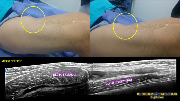
- Clinical and examination: A 35-year-old male patient presents a bulge in the proximal third and a depression in the distal third of the anterior compartment of the right thigh, soft, non-painful, without inflammatory changes. (Image 1). Background: 3 weeks ago, while playing soccer, he felt a pull when kicking the ball, fell to the ground unable to stand up, and then extensive bruising appeared.
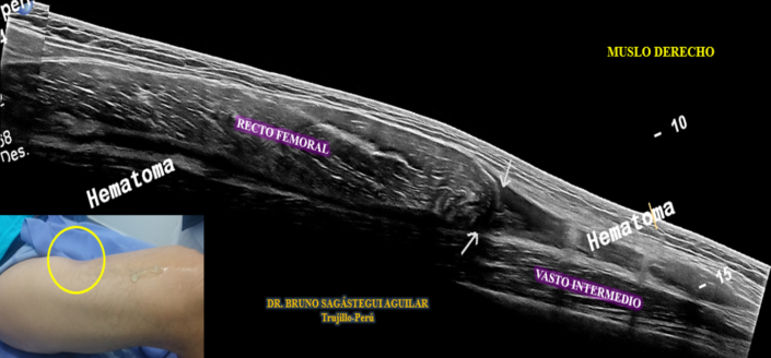
- Ultrasound findings: Retraction of the rectus femoris muscle towards the proximal (bell clapper) is observed due to rupture of the distal myotendinous junction. (Image 1 and 2), Anechogenic collection is associated in the lateral and lower portion (hematoma) that separates it from the deep fascia. (Videos 1 and 2)
- Diagnosis: The findings are compatible with a complete tear of the distal myotendinous junction or posterior fascia of the right rectus femoris.


