Spigelian hernia in the right lower quadrant
Dr. Bruno Sagastegui Aguilar
- Case name: Spigelian hernia in the right lower quadrant.
- Author's name: Dr. Bruno Sagástegui Aguilar.
- Author's job position: Work center: SONOSCAN Ultrasound Center (Private) and TOMONORTE Diagnostic Imaging Center (Private) Trujillo – PERU.
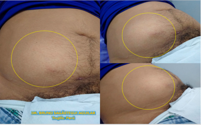
- Clinical and examination: Male patient, 42 years old, comes due to a “mass” in the lower right quadrant of the abdomen (Image 1) for 2 years, which increases in size when lifting weights or doing intense exercise and decreases when resting, causing pain. History: He denies surgery or trauma.
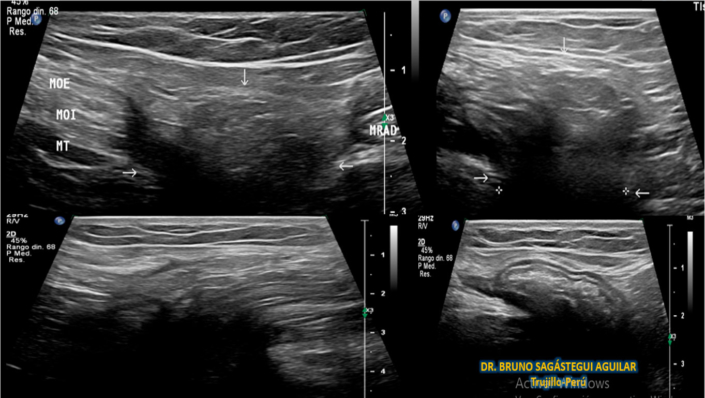
- Ultrasound findings: In the lower right quadrant, a hernial defect is seen between the lateral border of the rectus abdominis muscle and the semilunar line. (Image 2), by which a large hernial sac protrudes through the aponeurosis of the transverse muscle, inside the aponeurosis of the external oblique muscle, containing mesenteric fat and intestinal loops, during the Valsalva maneuver. It is partially reduced by echo pressure. (Image 2, Video 1 and 2).
- Diagnosis: The findings are consistent with Spigelian hernia in the right lower quadrant.


