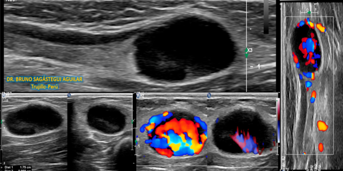
Left epitrochlear lymphadenopathy with inflammatory characteristics
Dr. Bruno Sagastegui Aguilar
- Case name: Left epitrochlear lymphadenopathy with inflammatory characteristics (due to cat scratch)
- Author's name: Dr. Bruno Sagástegui Aguilar.
- Author's job position: Work center: SONOSCAN Ultrasound Center (Private) and TOMONORTE Diagnostic Imaging Center (Private) Trujillo – PERU.
- Clinical and examination: Female patient, 3 years old, is referred by a pediatrician for a “bulge” in the distal medial region of the left arm (Image 1) near the epitrochlea, slightly painful to palpation with no external signs of inflammation. History: One week ago he had fever and increased volume in the area described above with moderate pain. He denies trauma. He raises cats at home.
- Ultrasound findings: An enlarged lymph node with well-defined edges with an eccentric hilar pattern and hypoechoic asymmetric thickening of the cortex is observed, measuring 17.5 x 9.5 x 11.8 mm; volume: 1.04 cc, seen in sagittal and axial views. (Image 2, Videos 1 and 2), shows color Doppler and CPA study increased hiliocortical vascularization (Image 2 and Video 3). In addition, an increase in echogenicity of the adjacent subcutaneous tissue is observed (Image 2, Videos 1 and 2) and dilation of the lymphatic duct with a course towards the forearm (Image 2 and Video 1).
- Diagnosis: Findings are consistent with left epitrochlear lymphadenopathy with inflammatory characteristics (due to cat scratch)


